


SOHLA : Mapping of microphytobenthos by hyperspectral imaging
Biomass mapping of microphytobenthos
A laboratory approach has helped develop the first radiative transfer model of the microphytobenthic biofilm : the MPBOM (Kazemipour and al., 2011). Associated with use of specific clues to discriminate micro and macro - algae on the mudflat , and in the biofilm of Euglena and Diatoms (Méléder and al., 2010; Kazemipour, 2011), this model was successfully applied to the 2009 Bay of Bourgneuf images (Kazemipour and al., 2012). The zero state monitoring is given as from the 2009 year. The campaigns of 2010 and 2011 gives us a first look at the evolution of microphytobenthos of the Bay of Bourgneuf . It is sharply down in 2010 compared to 2009 but the weather conditions were not favorable .
Material
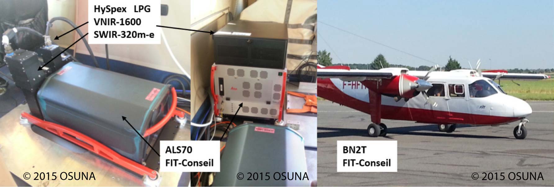
The 2002 image was produced by DLR with its DAIS camera through HySens program. The other images of the airborne SOHLA program have been conducted since 2009 by Actimar and FIT-Conseil, using cameras HySpex VNIR-1600 and SWIR-320m-e of LPG financed by CPER Génie-civil environnemental et développement durable de la ville (axis 3).
Here is the material used to achieve the 2013 images:
Studies in the Bay of Bourgneuf
- RGB images of the Bay of Bourgneuf :
financed by HySens 08/2002 give to GEOPAL  Click image to enlarge it | financed by GERRICO+CNRS 09/2009 give to GEOPAL  Click image to enlarge it | financed by CNRS 05/2010 give to GEOPAL 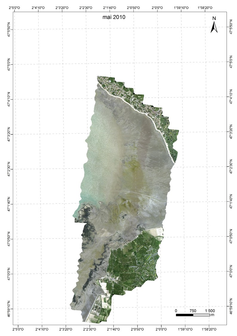 Click image to enlarge it |
financed by GEOPAL 09/2010 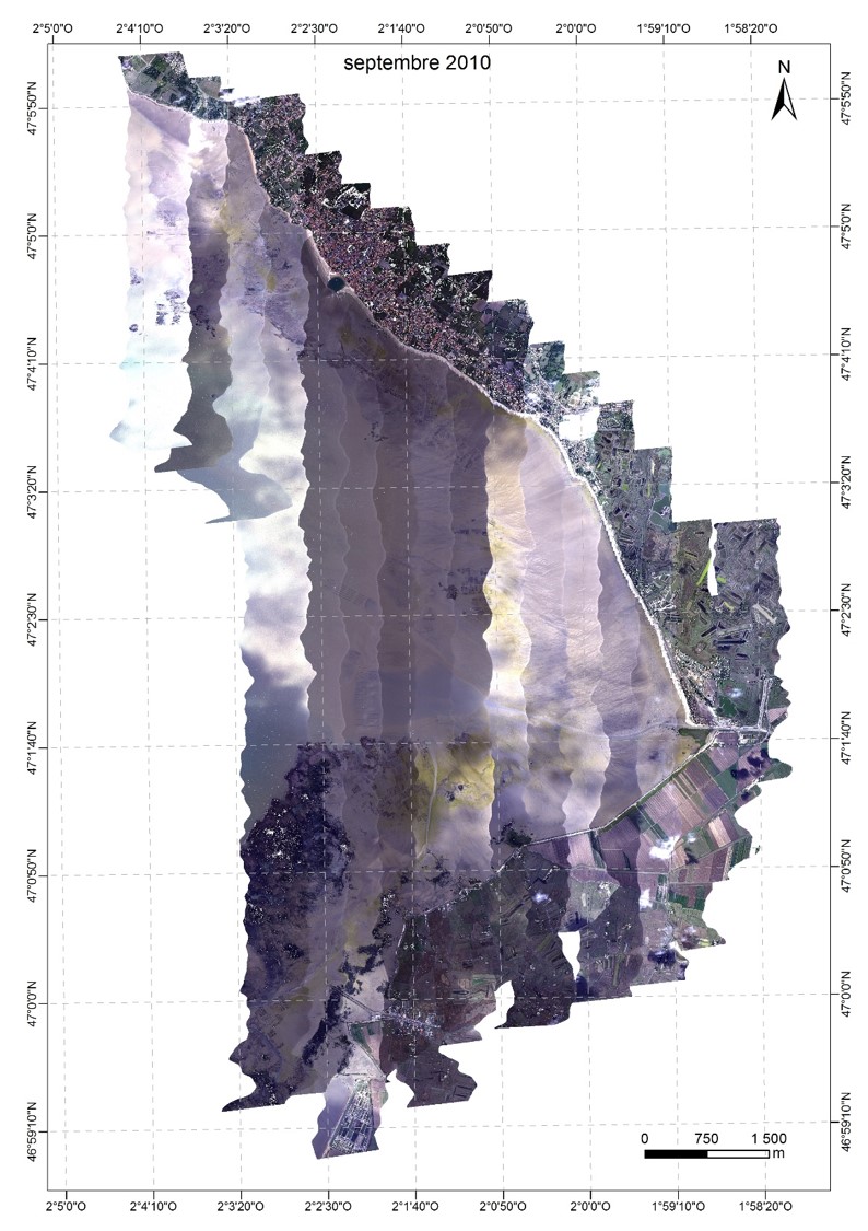 Click image to enlarge it | financed by GEOPAL 09/2011 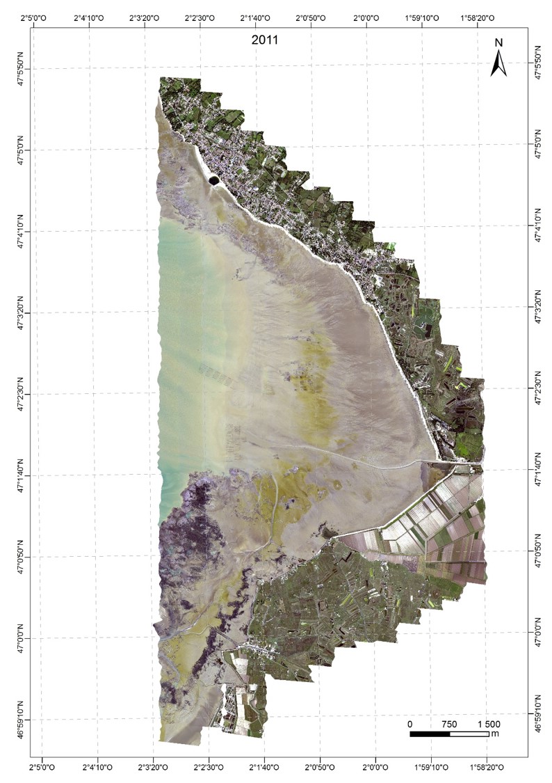 Click image to enlarge it | financed by GEOPAL 09/2013 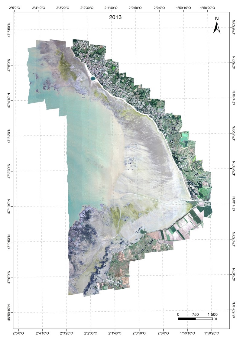 Click image to enlarge it |
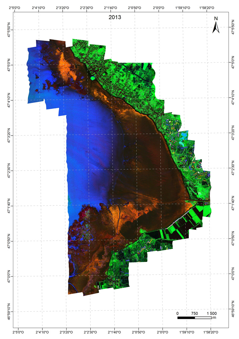
- Images obtained after analysis of previous images, highlight red-orange diatoms , green euglena (with terrestrial vegetation ) and in blue the presence of a clear water layer on the surface by the combination of specific indices each of the covers. The most turbid waters appear black like other materials.
| Image after image acquired in 2002 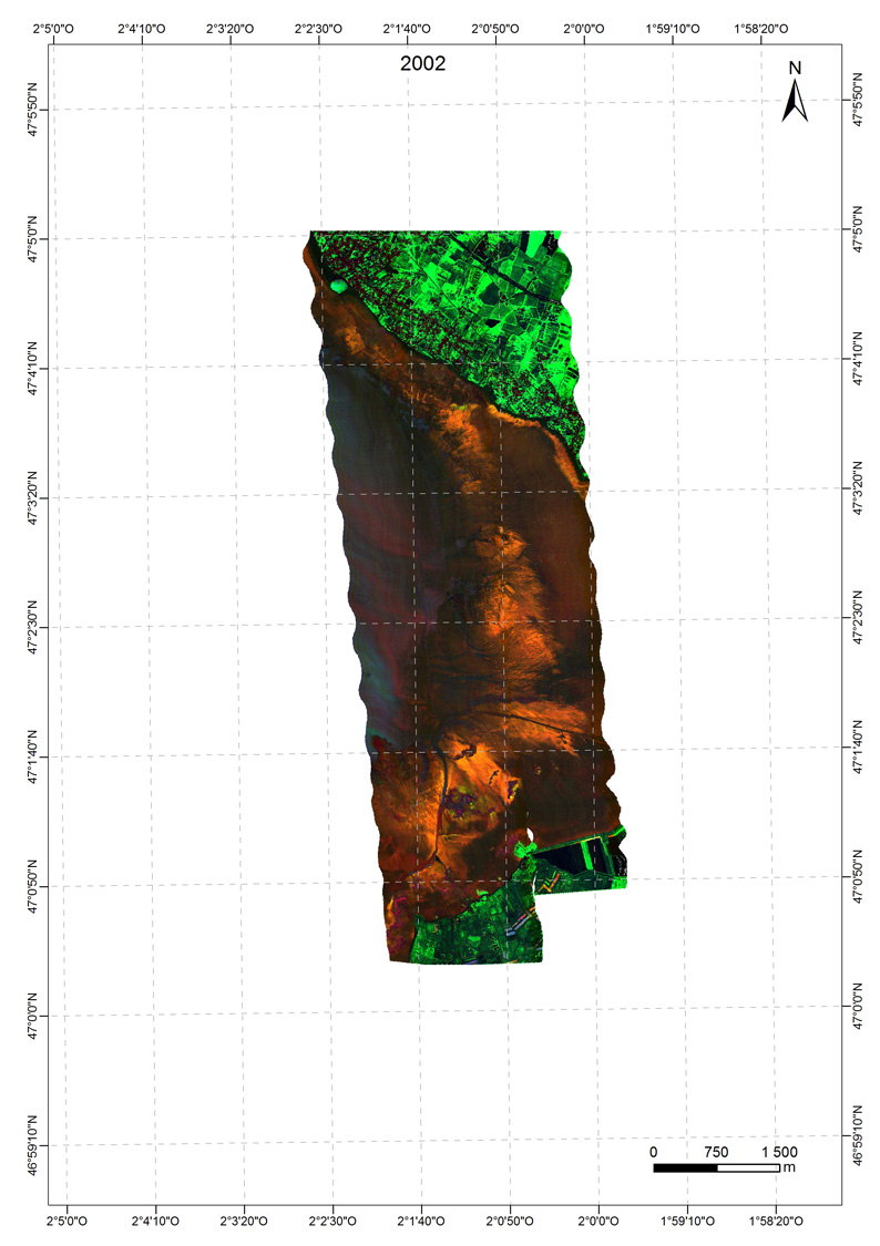 Click image to enlarge it | Image after image acquired in 2009 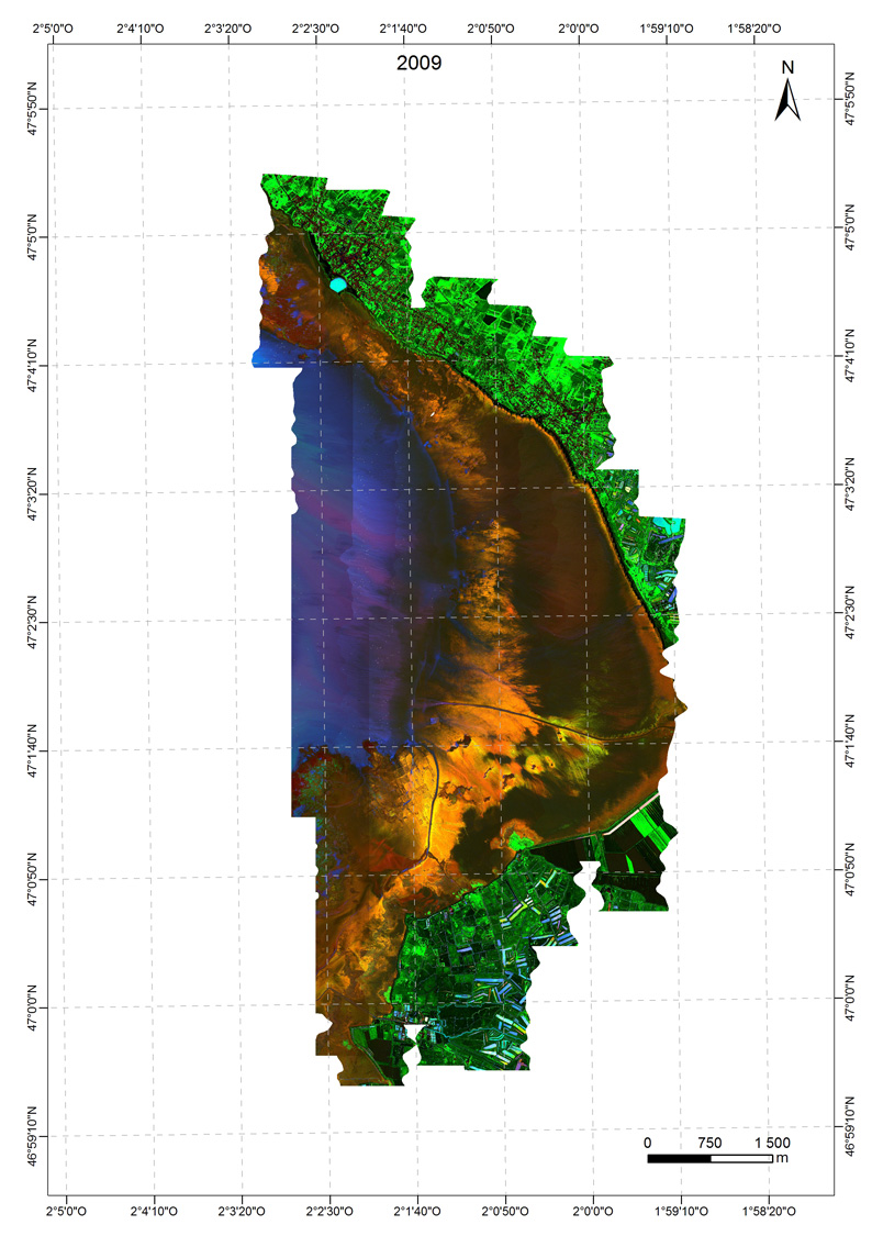 Click image to enlarge it | Image after image acquired in may 2010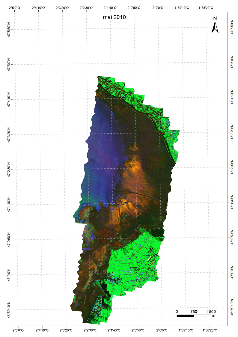 Click image to enlarge it |
Image after image acquired in september 2010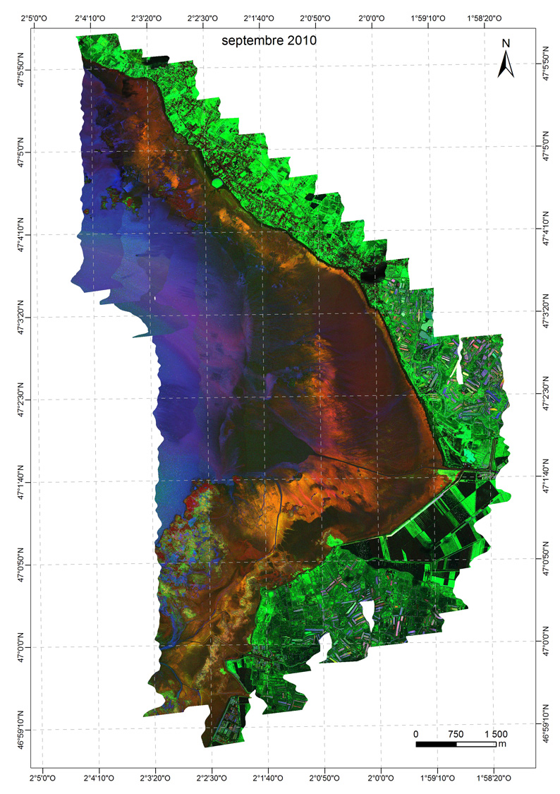 Click image to enlarge it | Image after image acquired in 2011 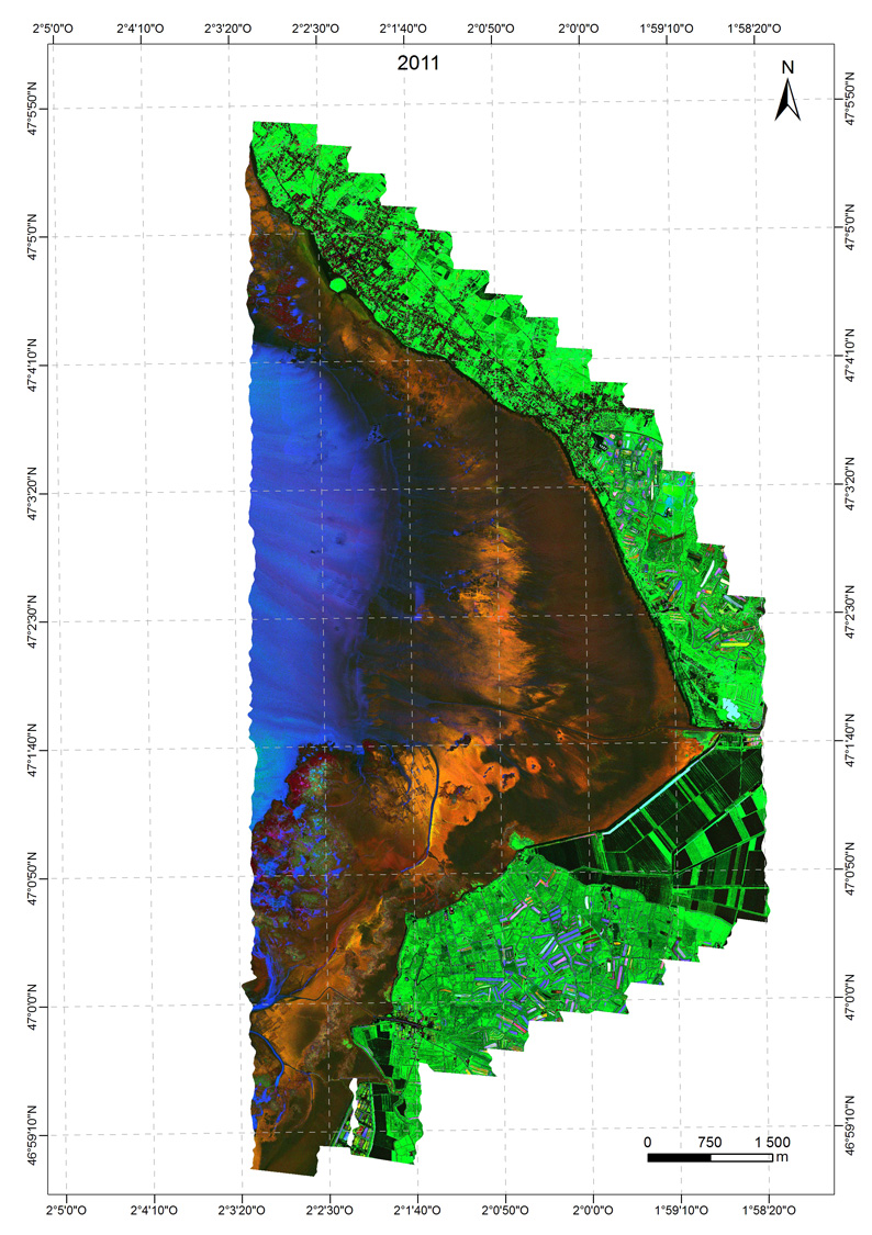 Click image to enlarge it | Image after image acquired in 2013 |
- Mapping of microphytobenthos by determining its biomass obtained after analysis of previous images by MPBOM:
Map images acquired in 2002 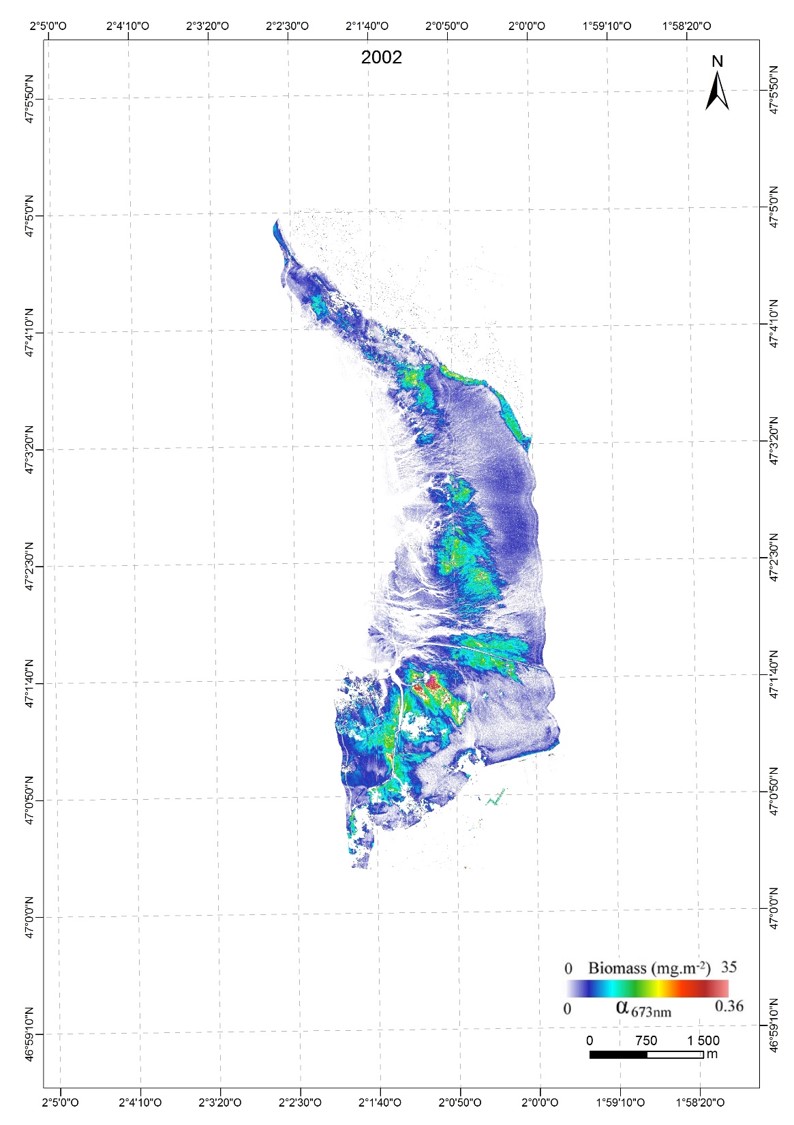 Click image to enlarge it | Map images acquired in 2009 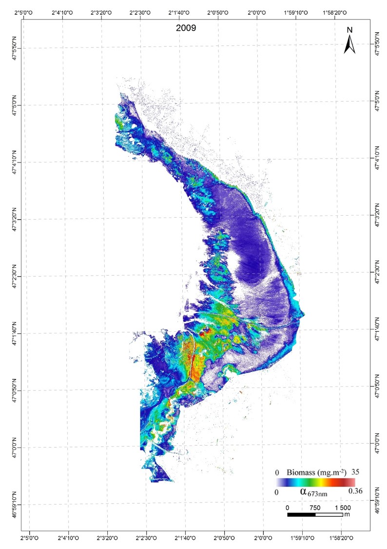 Click image to enlarge it | Map images acquired in may 2010 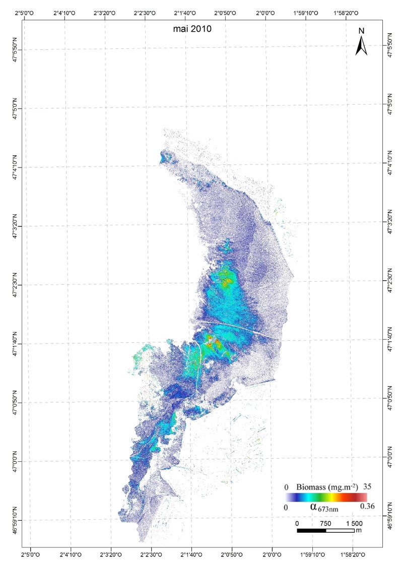 Click image to enlarge it |
Map images acquired in september 2010 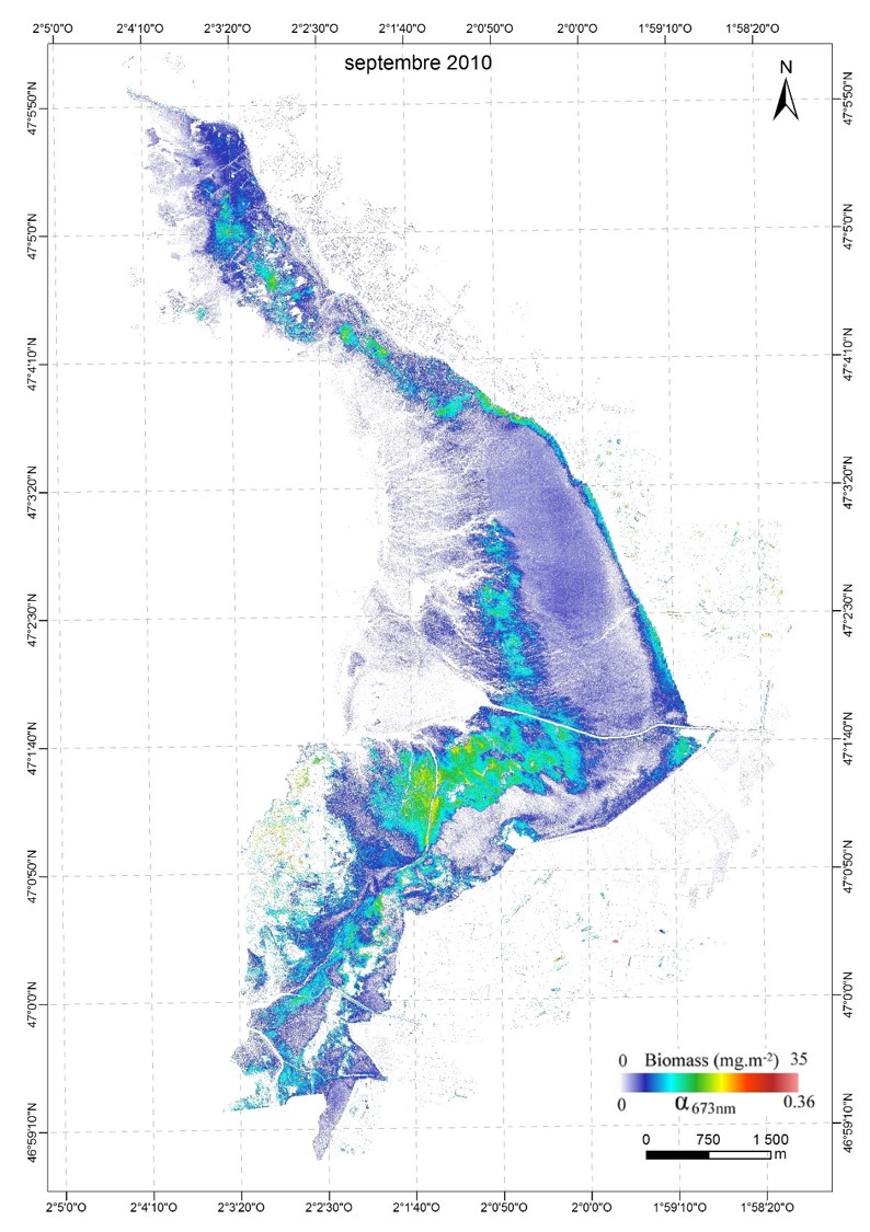 Click image to enlarge it | Map images acquired in september 2011 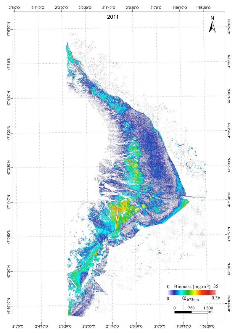 Click image to enlarge it | Map images acquired in september 2013  Click image to enlarge it |
The same procedure was applied to the images on the estuary of the Loire , to highlight the zonation of themicrophytobenthos.
Studies on the Loire Estuary
- RGB images of the estuary of the Loire
made by ACTIMAR in 2010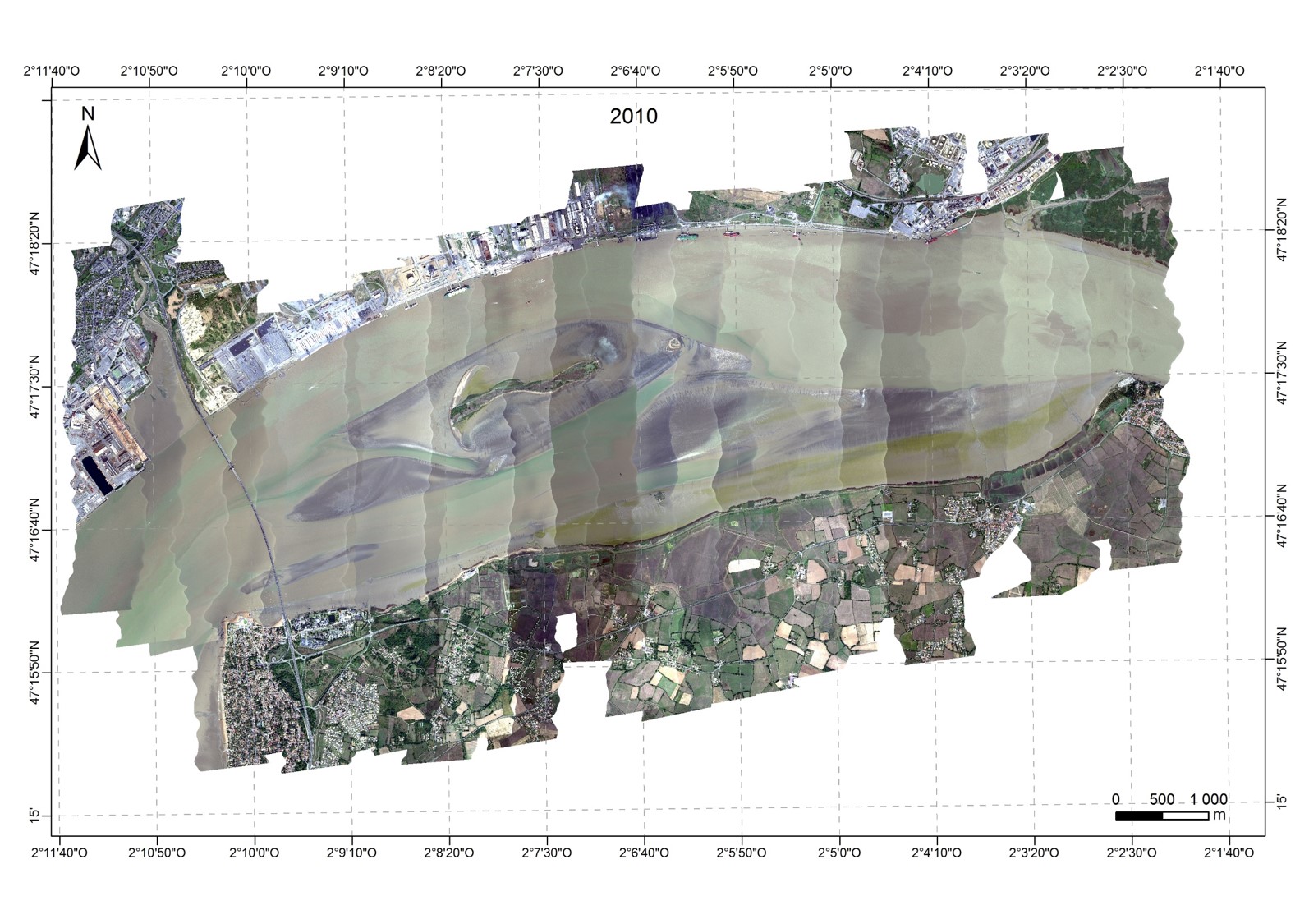 Click image to enlarge it | made by ACTIMAR in 2011 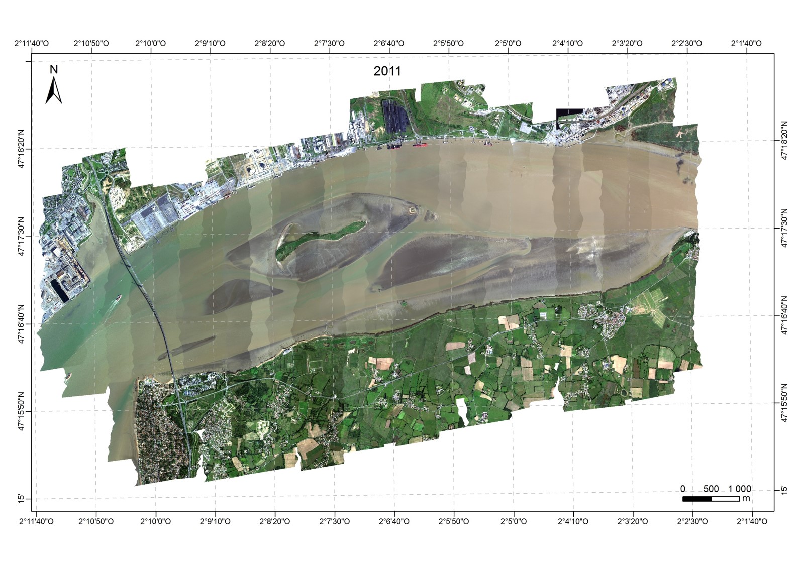 Click image to enlarge it | made by FIT-Conseil (with reduction of flight time) in 2013  Click image to enlarge it |
- Images obtained after analysis of previous images, highlight red-orange diatoms , green euglena (with terrestrial vegetation ) and in blue the presence of a clear water layer on the surface by the combination of specific indices each of the covers. The most turbid waters appear black like other materials.
images created in 2010 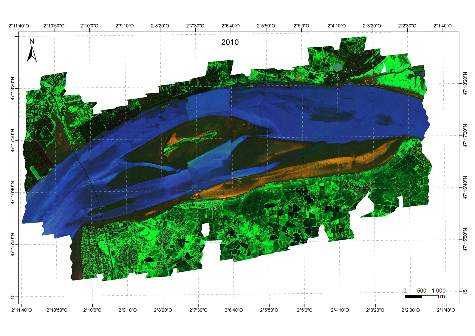 Click image to enlarge it | images created in2011 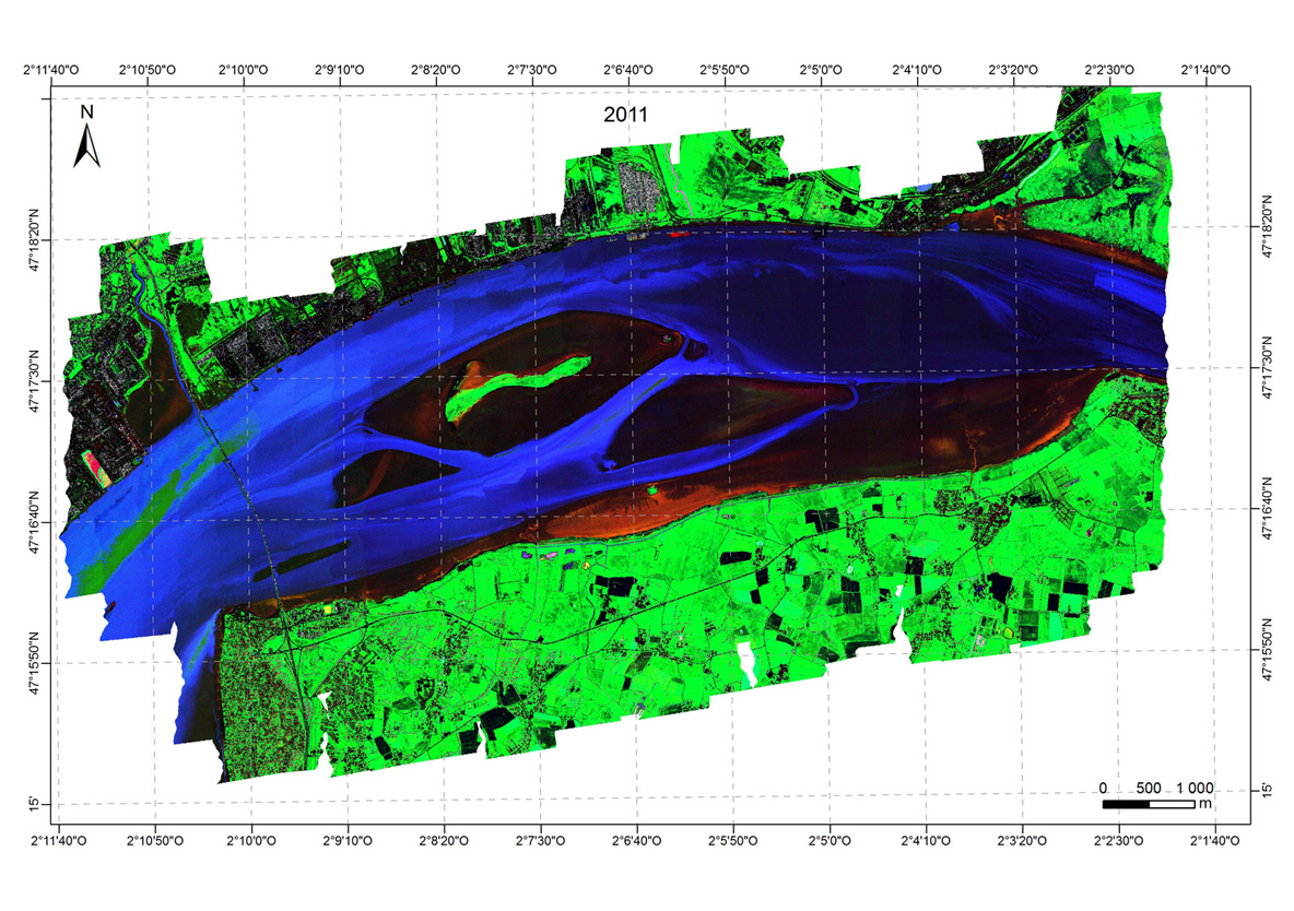 Click image to enlarge it | images created in 2013  Click image to enlarge it |
- Mapping of microphytobenthos by determining its biomass obtained after analysis of previous images by MPBOM::
Map images acquired in 2010  Click image to enlarge it | Map images acquired in 2011 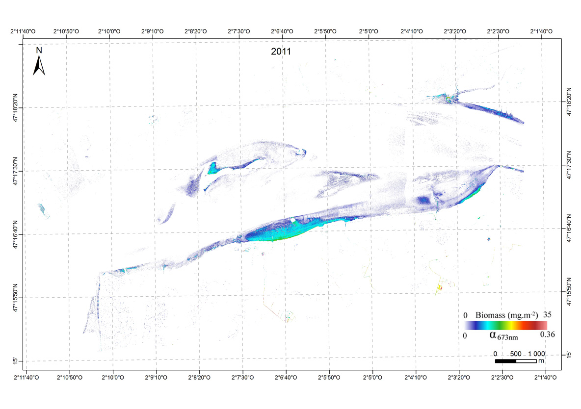 Click image to enlarge it | Map images acquired in 2013  Click image to enlarge it |
The main results are that the microphytobenthos varies in the same range of biomass, with maximum values of 35 mg Chl a/m². While the Bay of Bourgneuf shows a fairly permanent structure of biofilms, they have a variable spatial distribution on the mudflats of the Loire estuary for three years studied. This heterogeneity is explained by a greater hydrodynamics in the estuary. These results are of utmost importance for understanding the functioning of these ecosystems with high productivity, and under strong anthropogenic influence.
Availability of data
Metadata are available in the metadata catalog of OSUNA.
They are also available in the SDI GEOPAL of the region of Pays de la Loire (bottom right of the page in the category catalog harvested, click OSUNA).
The data are available online from the map viewer OSUNA, using the map viewer INDIGEO. A mappping project has been created specifically for this work (for more information on the data sets please contact the authors).
Bibliography
Kazemipour, F., (2011) Caractérisation hyperspectrale des biofilms microphytobenthiques : Cartographie de la biomasse de la micro à la macro échelle. Thèse de Doctorat de Nantes Université. 198 p.
Kazemipour, F., P. Launeau, V. Méléder (2012) Microphytobenthos biomass mapping using the optical model of diatom biofilms: Application to hyperspectral images of Bourgneuf Bay. Remote Sensing of Environment 127, 1-13
Kazemipour F., V. Méléder, P. Launeau (2011) Optical properties of microphytobenthic biofilms (MPBOM): Biomass retrieval implication. Journal of Quantitative Spectroscopy and Radiative Transfer, Volume 112, Issue 1, January 2011, Pages 131-142
Méléder V., Launeau P., Barillé L., Combe J.-Ph., Carrère V., Jesus B., Verpoorter Ch. (2010). Hyperspectral imaging for mapping microphytobenthis in coastal areas. In : Geomatic Solutions For Coastal Environments, Eds Maanan M. and Robin M. Nova Science Publishers, Inc. pp. 71-139
 |  |
 | |  |
Mis à jour le 07 December 2015.